The microscope is the new drill
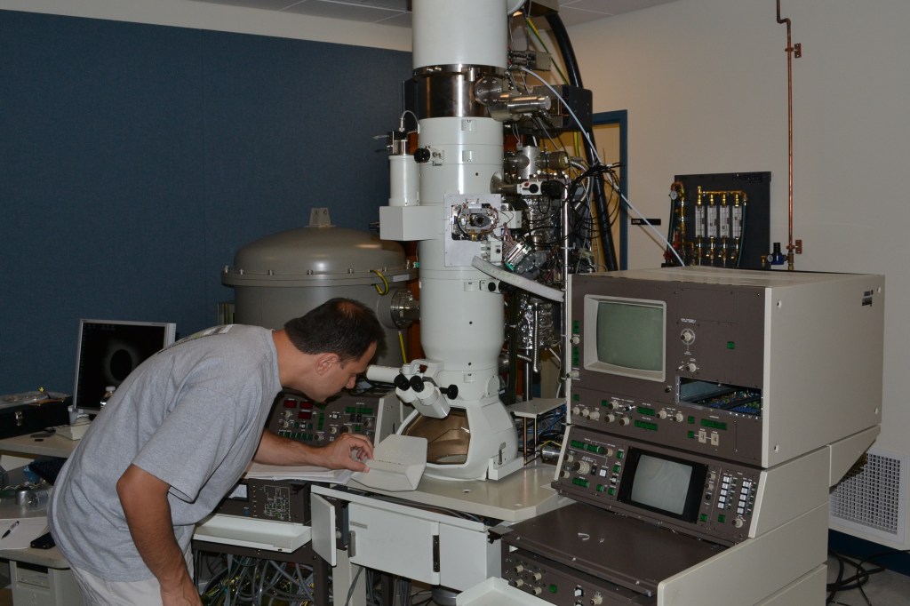

Photo by Lori Lennon.
How many things can you do with a microscope? Before this morning I would have probably said “one: magnify stuff.” Now I can confidently say “two: magnify and drill holes in stuff.”
Meni Wanunu, assistant professor of physics and chemistry/chemical biology, will be using the university’s new transmission electron microscope (TEM) to make nanopores nucleic acid analysis and DNA sequencing.
TEM differs from a standard light-microscope in that it uses a beam of electrons as the “illumination” source instead of a beam of photons. A series of electromagnets work to focus the electron beam before and after puncturing the material. For imaging the material, the beam needs to be relatively diffuse, said Wanunu. Otherwise it would destroy the sample.
Wanunu’s work in nanopores takes advantage of that exact drawback. Instead of using a diffuse electron beam, he focuses the beam down to a couple of nanometers. At this scale, the beam is incredibly precise and powerful and can drill a clean, exacting hole into a membrane. Some materials Wanunu has already drilled include silicon nitride and single-layer graphene membranes.
The original nanopore technique used ion beams instead of electrons, but using electrons rather than ions offers much improved precision and less sample contamination.
While the instrument was purchased to enable Wanunu’s work, it will also be available for other researchers interested in drilling tiny holes or magnifying things. It is capable of making things only a couple Angstroms wide visible to the naked eye. That’s the size of a single atom.
But doing so requires a lot of power. The electron beam alone comes from a huge 200K volt battery!