Killing cancer the simple way
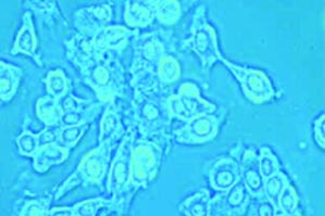

The nanocarriers in a mixture of cancerous and healthy cells
Targeted drug delivery is a hot topic these days. Chemotherapy, for example, blindly kills anything in its path — these drugs don’t distinguish between healthy cells and cancerous cells; they just kill cells. Period.
Professor Vladimir Torchilin and his buddies at the Center for Cancer Nanotechnology Excellence are developing nanoscale drug delivery technologies, which I like to think of as microscopic Trojan horses. Very basically, the outside is covered with proteins that make it tumor friendly while the inside contains a deadly payload that is unleashed after the cancer cell welcomes it through its gates. Healthy cells don’t recognize the surface proteins and thus don’t get the chance to be exposed to the drug.
This is a great idea of course, but as you might imaging, making microscopic Trojan horses, or nanocarriers, as they’re called, can be rather challenging. At this year’s RISE:2012 conference I met a graduate student in Torchilin’s lab, Tao Wang, who has developed a simple, straightforward approach that could take targeted drug delivery from “hot topic” to “hot practice.”
Getting the surface proteins onto these nanocarriers is often costly and chemically complicated (how’s that for alliteration!?) Wang’s approach overcomes these problem by letting biology do all the hard work.
Bacteriophages are viruses that infect bacterial cells by lodging themselves in the cell membrane and depositing genetic material inside which messes up the bacterium’s standard operating procedures, so to speak. A process called “phage display” developed ’80s allows scientists to tag the ends of bacteriophages with various protein fragments.
There’s a convenient similarity between bacterial cell membranes and nanocarriers: they are both made out of lipids, which are hydrophobic (water hating) molecules. Bacteriophage proteinss are also hydrophobic, which is what allows them to buddy up to the bacterial cell membrane. The protein fragments tagged to its end, however, is hydrophilic, which means it will want to hang out on the outside of any lipid membrane it finds itself near.
Do you see where this is going yet? The group sifted through a huge library of protein-tagged bacteriophages until they found one that was recognized by breast cancer cells. Then they mixed the phage-derived protein with a the nanocarrier containing a drug that kills breast cancer cells. The bacteriophage lodged itself inside the nanocarrier’s lipid membrane, as if it were a bacterial membrane, with the protein fragment dangling on the outside. Then they mixed these little guys with cancerous and non cancerous cells in a test tube and found that they attacked cancer cells and bypassed healthy cells.

A foregin gene inserted into the bacteriophage (left) tags it with a protein fragment, which is recognized by receptors on the surface of a cancer cell but not a healthy cell. The tagged bacteriophage spontaneously nestles into the lipid membrane of the drug-containing nanocarrier (right).
All this work was published in a paper in 2010 in the journal Nanomedicine. Wang’s poster at RISE:2012 illustrated some of the work she’s been doing since then, using this technique to cure cancer in mice.
First she had to give the mice cancer, which I’m sure wasn’t good for the moral compass, but then she had the satisfaction of knowing that her drugs actually helped them.
When she gave them the drug inside a nanocarrier that didn’t have the bacteriophage fusion protein incorporated, they showed very little reduction in tumor size.
But when she delivered the drug via bacteriophage-nanocarriers new tumor growth slowed down and existing tumor size reduced significantly. The mice maintained healthy body weights and “showed no signs of discomfort.”