Northeastern researchers find a faster and more sensitive way to study proteins, which could lead to advances in disease treatment

Protein complexes are important for the majority of vital processes in the cell and human body, such as producing energy, copying DNA and regulating the immune system.
Composed of groups of connected protein chains called subunits, the complexes are also good targets for medicines that treat diseases.
But studying them in their native, natural physiological state, while preserving their 3-D protein folds, has proved challenging.
Traditional mass spectrometry methods and structural biology techniques may require breaking protein chains into pieces or turning protein parts into crystals.
These approaches not only disrupt the structure of the assembled protein molecules but involve using substantial amounts of samples and waiting weeks for results.
Now researchers at Northeastern University have developed a novel method of preserving the structure of protein complexes and their interactions under near-native conditions while analyzing them in 30 minutes or less, using small sample amounts.
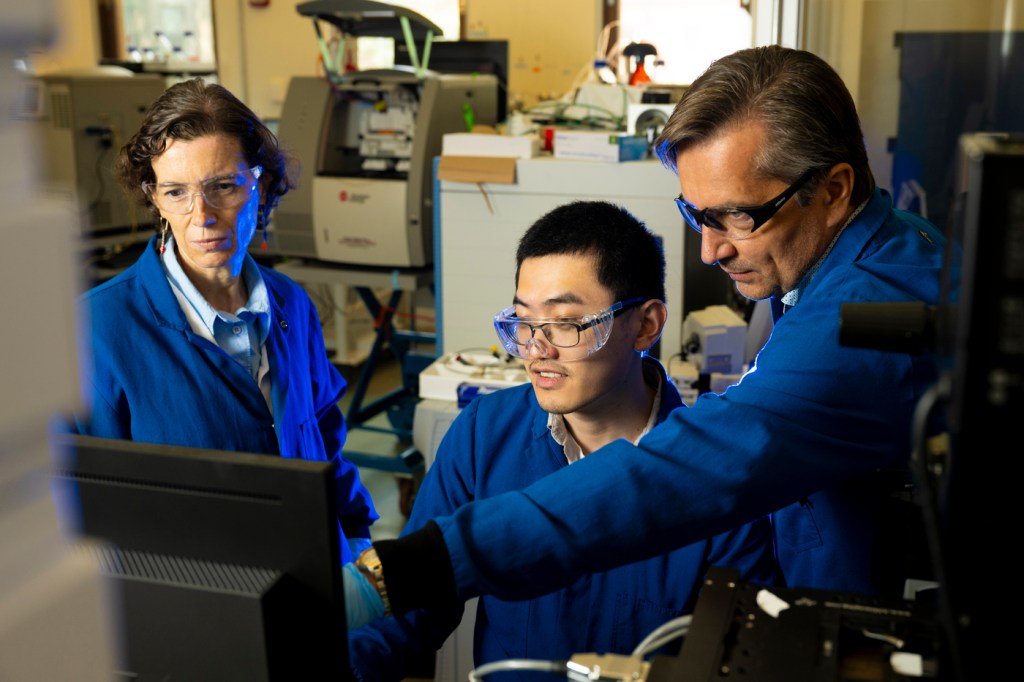
Associate research scientist Anne-Lise Marie and associate professor of chemistry and chemical biology Alexander R. Ivanov say their research, published in the Advanced Science journal, could eventually expedite drug development for pathologies such as Alzheimer’s and Parkinson’s disease.
Their method uses a research technique called capillary electrophoresis-mass spectrometry (CE-MS) to study the conformational changes of proteins and protein complexes
The researchers report it is fast and highly sensitive. “Our method substantially minimizes sample consumption and sample loss, simplifies and shortens the analytical workflow” and maintains the protein under study at “near-physiological conditions,” they say.
“The whole direction is pretty novel,” Ivanov says.
“The main advance here is to show that capillary electrophoresis coupled to mass spectrometry can enable a structural analysis of big protein complexes, which are essential in conducting many biological functions,” he says.
“The vast majority of biological reactions and functions are enabled by protein complexes,” Ivanov says.


“Changes in the conformation of a protein may lead to structural destabilization, aggregation and loss of biological activity, which may result in devastating human pathologies, including neurodegenerative and oncological diseases in humans,” he says.
“Because of this, it is important to develop analytical techniques that are capable of detecting, characterizing, and monitoring structural changes of proteins and their interactions with other molecules (small and large) in real time and in the liquid phase, to mimic what happens in vivo,” Marie says.
Proteins and protein complexes in their natural, native state, she says, are “better representatives of biological systems.”
Mass spectrometry uses ion detection systems and other tools to identify and quantify different molecules in a biological or clinical sample.
The addition of capillary electrophoresis also allows researchers to efficiently separate molecules, including proteins, carbohydrates and nucleotides, by immersing them in a solution and drawing them into a glass capillary less than a human hair in diameter.
This enables their separation based on the molecule’s charge and size under high voltage prior to mass spectrometry analysis, allowing the researchers to observe them interacting with other molecular species.
Editor’s Picks
In this study, Marie and Ivanov explored interactions of a large protein complex with nucleotides, metal ions, protein substrates and other protein complexes.
“We were also able to pinpoint the mutations, the changes in the sequence of amino acids that we specifically introduced,” Ivanov says.
“Many diseases are caused by mutations. Here we show that we can actually start with a gigantic, native protein complex and go all the way down to characterization of its primary structure and find minor protein sequence perturbations, down to point mutations,” he says.
Marie says the intention of the developed native CE-MS technique, which is at the interface of analytical chemistry, proteomics, and structural biology, is not “to compete with conventional structural biology techniques like X-ray crystallography or cryo-electron microscopy.”
“We think we developed a relatively high throughput, robust, and efficient complementary technique, which is extremely sensitive,” she says. “The required amounts of proteins are about 10,000-fold lower compared to conventional biochemical/biophysical techniques.”
The sample amounts are equivalent to approximately 200 to 300 small cells, “compared to many millions and billions required in conventional studies,” Ivanov says.
“Also, conventional structural biology techniques can take weeks to get results while CE-MS analyses can take less than 30 minutes,” Marie says, adding that the new approach “enabled us to follow the dynamic changes of proteins in solution and in real-time.”
“The method could help investigate the interactions between potential drug candidates and biologically critical proteins, for the detection, investigation, monitoring, and treatment of human pathologies, using minute sample amounts,” Marie says.
“Broadly speaking, the method could help answer a myriad of questions in biomedical and clinical or fundamental biology applications,” Ivanov says.


