Breakthrough 3D imaging to detect lung cancer early developed by Northeastern researcher
Photoacoustic imaging technology (PAIL) for diagnostic lung assessment is being developed by Soner Sonmezoglu, the principal investigator for the project funded by an award from the ARPA-H.

A Northeastern University researcher is developing new imaging techniques that would allow physicians to detect lung cancer earlier than ever.
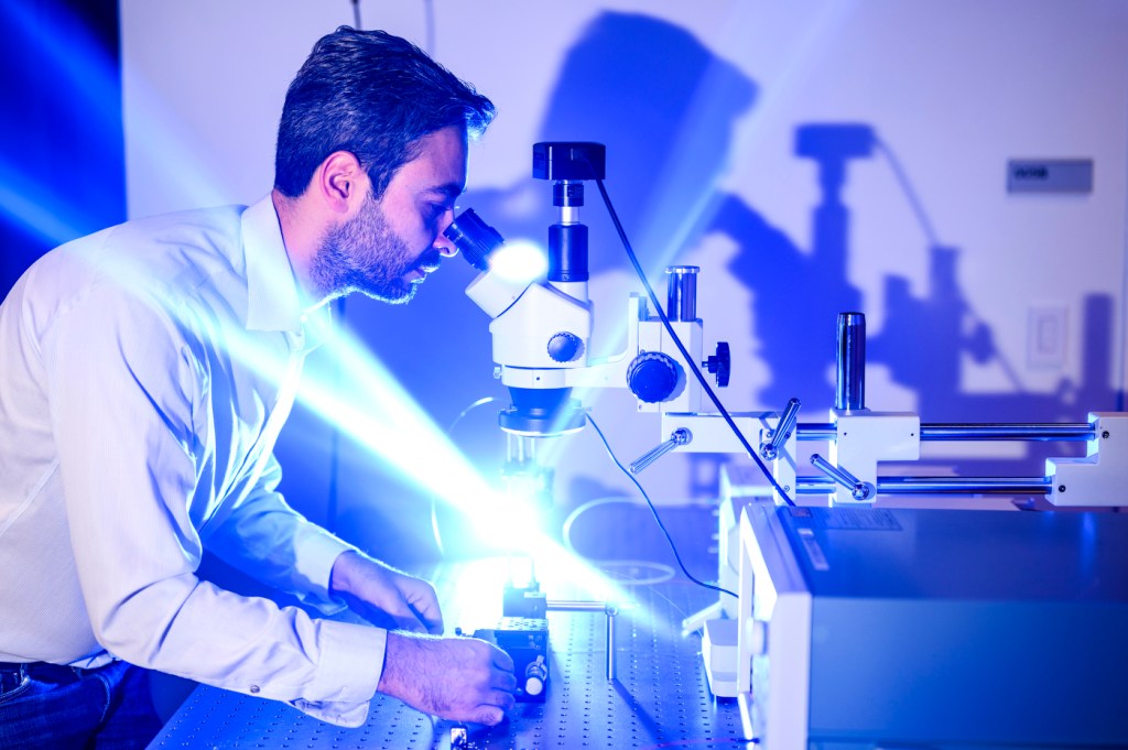
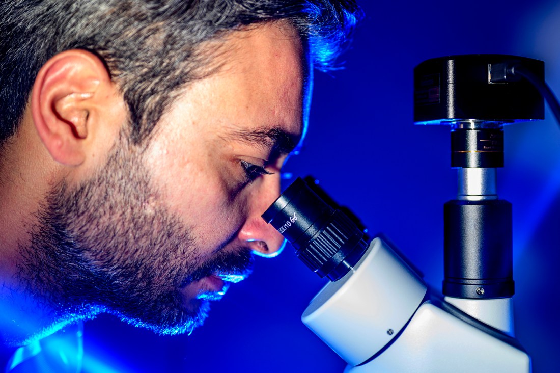
Soner Sonmezoglu is leading a multi-institutional team that will develop a system that uses a miniaturized probe the size of a grain of rice and high-resolution, 3D imagery to recognize early-stage cancer before it grows and spreads.

“We are building a technology at Northeastern to identify the tumors if they are malignant or benign when they are small,” says Sonmezoglu, Northeastern assistant professor of electrical and computer engineering.
Lung cancer is the leading cause of cancer death in the U.S., accounting for one in five cancer deaths, according to the American Cancer Society, which says that patients whose malignancies are treated before they spread to other parts of the body have a higher five-year survival rate.
The problem is that current imaging techniques do not have high enough resolution to determine whether tumors in their early stages are malignant or benign without the aid of a biopsy, says Sonmezoglu, the principal investigator for the project funded by an award up to $13.2 million from the Advanced Research Projects Agency for Health.
And “biopsies have a lot of complications,” he says. “They can cause the lung to collapse,” among other things.
“They also put a financial burden on the healthcare system because a biopsy is an expensive procedure,” Sonmezoglu says.
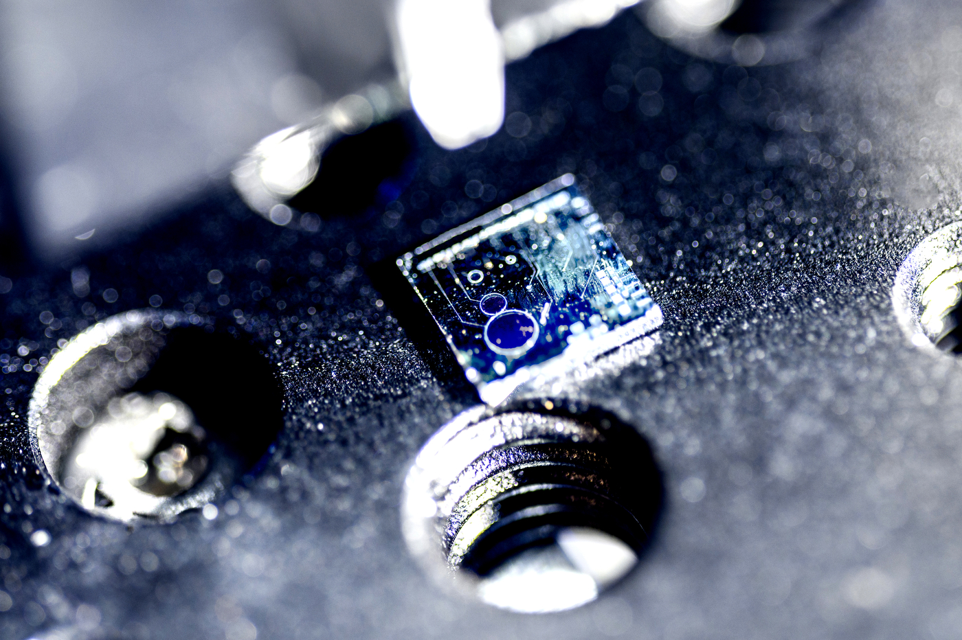
The system being developed at Northeastern will use a disposable probe that is 1.5 millimeters in diameter and a reusable electronic box to gather high-resolution, 3D images that convey functional and structural information about tumors.
It is called an electronic photonic photoacoustic imaging system and will be the first of its kind, says Sonmezoglu, an expert in integrated circuits and microfabrication, which use photons instead of electrons to process and distribute information.
“This technology enables us to miniaturize the probe that touches the lung tissues through a bronchoscope,” he says. “It’s more than an order of magnitude smaller than the other probes that exist today.”
The system will use smaller images from cameras at higher speeds and with less thermal effects.
“You can identify really tiny, small features, for example, the vascular map of a tumor and healthy tissue,” Sonmezoglu says. “It can also look at oxygen saturation and fibrosis,” which is often associated with cancer, he says.
“These are the features we are going to look to identify,” he says. Physicians will study them to identify which ones indicate whether the tumor is benign or malignant.
The miniaturized optical photoacoustic imaging system being built by Northeastern’s team will employ the use of advanced image reconstruction algorithms, Sonmezoglu say.
He says that while lung cancer will be the focus of the proof-of-concept demonstration during the five-year program, the commercialization of this imaging technology will have broad societal impacts.
It can provide ample opportunities to improve diagnosis, personalized treatment plans, and, eventually, patient outcomes.
Collaborating with Northeastern University are pathologists, surgeons, and engineers from Massachusetts General Hospital, Johns Hopkins University, and the University of Washington.
“The medical doctors that I reached out to work on lung cancer and are already familiar with using technology with probes and bronchoscopes, and working with MGH and Johns Hopkins, we will have access to many medical doctors whose guidance and input will help in design and faster adoption in the clinical environment,” Sonmezoglu says.
He says if successful, the new imaging system would change the landscape of the photoacoustic imaging field by providing real-time and high-contrast 3D images that can help with earlier diagnosis of diseases.
“The system and technology can be applied to other cancer types beyond lung cancer, including prostate, ovarian, and bladder cancers,” Sonmezoglu says.