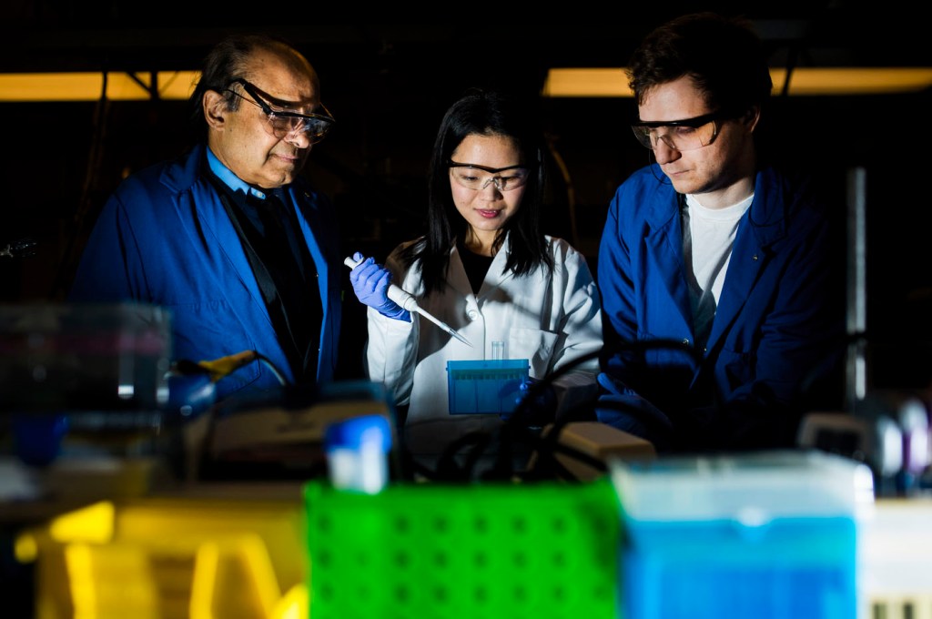
Professor on a mission to create crystal-clear images of the human brain

A groundbreaking MRI technique developed by Northeastern professor Srinivas Sridhar and his students uses magnetic nanoparticles to create detailed maps of blood vessels and blood volume in the human brain. These maps could allow doctors to more easily spot anomalies and blood-flow problems as well as diagnose and treat neurological disorders such as Alzheimer’s, multiple sclerosis, and traumatic brain injuries. A spinoff company founded by Sridhar, Theranano, recently received a $225,000 grant from the National Institute on Drug Abuse to test the technique in clinical trials at Massachusetts General Hospital.
“In my career, I’ve written more than 200 papers and gotten more than $20 million in grants. But I’ve wanted to make sure that the work benefits people. It’s very exciting that, only three years after my students and I began this research, it will be tested in a clinical setting,” says Sridhar, University Distinguished Professor of Physics, who worked with students Codi Gharagouzloo, PhD’16, Ju Qiao, PhD’18, and Liam Timms, PhD’18, on the breakthrough.
Traditional MRIs require that patients be injected with a liquid contrast agent so that their blood vessels are illuminated and easier to read on a scan. But the most commonly used agent is gadolinium, which can be toxic to organs. The technique created by Sridhar and his students deploys iron oxide nanoparticles, which are safer for the human body. The nanoparticles attach to blood flowing through the brain then light up under an MRI, forming intricate pictures.
The new technique has two special features, Sridhar says: It generates “beautiful images with unprecedented clarity,” and it measures blood volume instead of just capturing a picture of it.
“Current MRIs make an image, then a radiologist assesses that image with the naked eye. Our technique is an instrument; it measures the amount of blood in each vessel,” says Sridhar, who is also the director of Northeastern’s Electronic Materials Research Institute. This more precise data could help doctors detect abnormalities.
In the clinical trial, doctors at MGH will use Sridhar’s method to map the brains of healthy patients, creating a template or baseline for the look and volume of normal blood vessels. Then they’ll map the brains of patients with neurological disorders to compare and determine how an abnormal brain looks and functions.
Sridhar hopes to extend this MRI method to other parts of the body that are especially vulnerable to gadolinium’s toxic effects, such as the heart, lungs, and kidneys.
“It could help with diagnostics—What is normal or abnormal for this part of the body?—and it could also help with treatment,” he said. “You could monitor how treatment is progressing and whether a particular medicine is creating the right kind of results for a disorder. The possibilities are endless.”


