‘Unicorn’ shipworm could reveal clues about human medicine and bacterial infections
Northeastern professor Daniel Distel and his colleagues have discovered a dark slithering creature four feet long that dwells in the foul mud of a remote lagoon in the Philippines. They say studying the animal, a giant shipworm with pinkish siphons at one end and an eyeless head at the other, could add to our understanding of how bacteria cause infections and, in turn, how we might adapt to tolerate—and even benefit from—them.
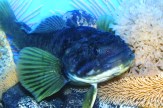
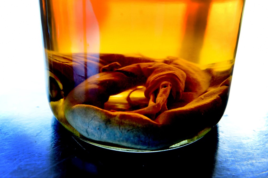
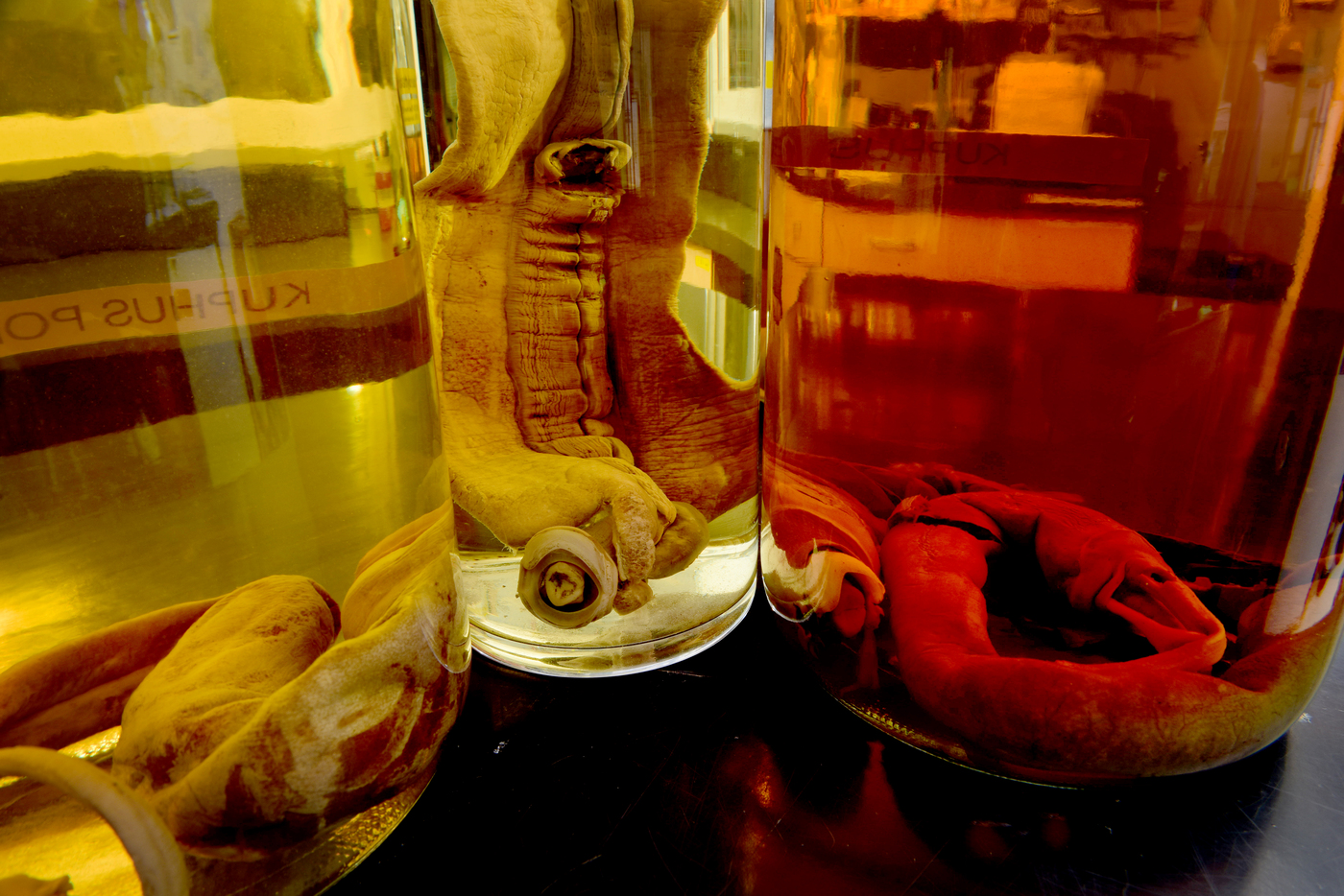
Live specimens of the massive shipworm, which was captured by Distel’s team with the help of researchers from the area, have eluded scientific description for hundreds of years. Distel, research professor at Northeastern’s Marine Science Center, has been searching for it for two decades. He had examined fragments of its tusk-like shell, made of calcium carbonate, and gazed wistfully at dead specimens preserved in ethanol. But neither he, nor any other living researchers, had ever come across a live specimen of the ancient species, a bivalve mollusc named Kuphus polythalamia that was first described (though incorrectly classified) by Swedish taxonomist Carl Linnaeus in 1758.
Now, in a new paper published in the Proceedings of the National Academy of Sciences, Distel and his colleagues present their research on the live shipworm. They describe how, remarkably, it does not eat, at least not much—it has a tiny digestive system—but instead bacteria living inside its gills convert sulfur gas from rotting wood into nutrients to keep it alive.
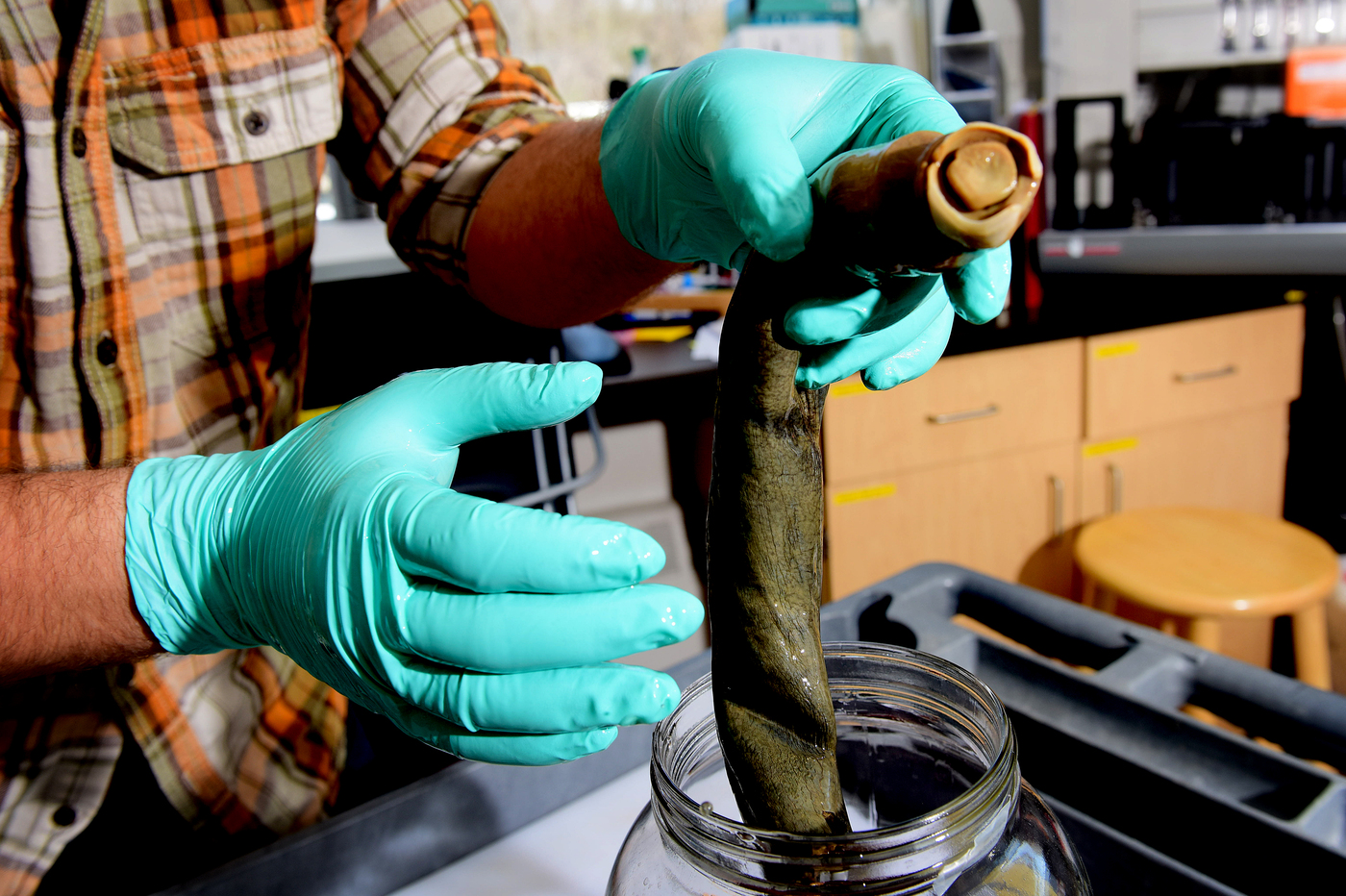
“Most shipworms are very delicate, translucent, usually white, beige or pink,” says Distel, who directs the Ocean Genome Legacy at Northeastern, a unique biological bank where researchers acquire DNA from organisms around the world for genetic analysis. “They’re mostly small, a few centimeters long. You have to be very careful not to damage them when you’re taking them out of the wood, where they live. This thing was like a baseball bat. It was a beefy, muscular animal, jet black.”
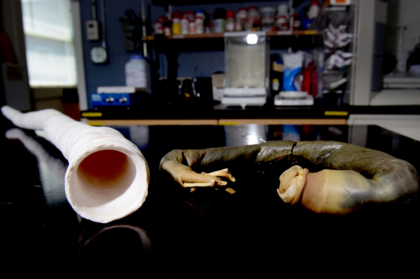
Indeed, the team had to edit much of the audio out of the original video of the creature’s debut. “When I took that thing out of the tube there was a collective gasp among the whole group,” says Distel, “along with quite a number of expletives that had to be deleted.”
An ‘evolutionary stepping stone’
Distel’s genomic analysis of the host Kuphus as well as the bacteria whipping up its food revealed a symbiotic relationship between the two that elucidates an “evolutionary stepping stone,” he says.
This thing was like a baseball bat. It was a beefy, muscular animal, jet black.
Daniel Distel
director, Ocean Genome Legacy
Shipworms as a rule eat wood; hence their being dubbed “termites of the sea.” Bacteria in their gills, Distel discovered earlier, secrete enzymes that travel to their gut and break down the wood—which is made of cellulose, an organic material—turning it into sugars.
But wood can also serve as a source of hydrogen sulfide—a sulfur gas that smells like rotten eggs. “We believe that somewhere along the line a shipworm acquired a sulfur-oxidizing bacteria as a symbiont, and it was able to get energy not just from the wood but also from the inorganic gas hydrogen sulfide coming from the wood as it rotted,” says Distel. “Eventually the new symbiosis completely replaced the old symbiosis.”
Other marine animals also get their nutrients from sulfur-oxidizing symbionts, but the sulfur source differs: The giant tubeworm Riftia pachyptila, for example, gets its sulfur from the effluence of volcanic hot springs on the sea floor.
The symbiont bacteria convert the hydrogen sulfide into food similar to the way photosynthesis works in green plants. Green plants take energy from sunlight and use it to synthesize sugars from carbon dioxide. The bacteria take chemical energy from hydrogen sulfide, pull carbon dioxide out of the seawater, and synthesize sugars and other nutrients.

Daniel Distel, director of the Ocean Genome Legacy at Northeastern, had been searching for the giant shipworm for two decades. Photo by Matthew Modoono/Northeastern University
“These bacteria live inside the animals’ cells, alongside the cytoplasm,” says Distel. “If we or any vertebrate had bacteria living inside our cells, we’d be very sick. In the long run, studying these symbioses may tell us a lot about the process of disease. What is it about these bacteria that they can infect the host yet not harm it? How does the host learn to tolerate, and even benefit from, the bacteria?”
Tracking a mystery
“For a biologist who is interested in these bivalves, it’s like a unicorn,” said Margo Haygood, a marine microbiologist at the University of Utah and senior author on the study, in a video revealing one of the giant shipworms.
Distel praises the power of social media for helping the researchers finally find the elusive giant.
They were in the Philippines in 2010 as part of a National Institutes of Health-funded project called the Philippine Mollusc Symbiont-International Cooperative Biodiversity Groups, studying molluscs and symbiotic bacteria in search of natural compounds that might be developed into antibiotics and other drugs. A student researcher reported seeing a YouTube documentary showing people on the island of Mindanao eating the shipworms, a delicacy, known in the area as Tamilok, that some believe has medicinal properties.
By the time the live creatures—packed in an odiferous brew of mud and seawater inside PVC pipes—arrived on the dissecting table at the University of the Philippines Manila, Distel’s head was spinning. “I was excited, amazed, and then concerned,” he says about seeing the animal slip out of its shell tubing after he’d cracked open one end. “What samples do we need to take? What tissues do we freeze? What tissues do we preserve in ethanol? What tissues do we preserve for electron microscopy? How do we preserve things for DNA studies? Remember, we didn’t know what we were going to get so we couldn’t fully prepare. Once we started cutting, amazement was gone and it was down to work, trying to figure out, ‘How do we not screw this up?’”