I’ll see your hydrogen and raise you a deuterium*

 Have you ever heard of hydrogen exchange as an analytical technique? I hadn’t until the day before yesterday. Actually, I hadn’t heard of hydrogen exchange, period. Forget the qualifier.
I know you’re bursting at the seams to find out, so I won’t make you wait any longer. Hydrogen exchange is exactly what it claims to be: the exchange of hydrogen atoms….
Okay. So what does that mean?
John Engen of the department of chemistry and the Barnett Institute has been working with this technique since his days as a PhD candidate and, more recently, has developed an analytical instrument that makes utilizing the process more approachable. Here’s how it works:
Proteins are big, lumbering molecules consisting of thousands of atoms. They are arranged in a well-defined sequence and then folded into what may seem like an amorphous ball of knotted-up yarn but is actually a very specific structure with pockets and helices perfectly situated for the protein to do its job. Hydrogen atoms decorate proteins throughout these intricate designs.
In your average Joe protein, hydrogen atoms have one proton and zero neutrons, giving them an atomic mass of 1. Deuterium is hydrogen’s black sheep brother, weighing in at 2 atomic mass units with a proton and a neutron.
Since deuterium and hydrogen are the same element with a similar atomic structure, they are interchangeable. Drown a protein in a bottle of deuterium oxide (aka heavy water or D2O), and the hydrogens on the surface will be replaced by deuterium, which is now in much higher supply. Once a protein is covered with deuterium atoms instead of hydrogen, the mass will increase significantly.
And this, my friend, is where the analytical piece comes in. Say you have a regular protein and a protein from a cancerous cell and you want to know what is different about the two. Submerge each in D2O and then weigh them with a “mass spectrometer” (a complicated molecular scale).
Think back to our knotted-up yarn ball. The cancerous yarn ball might be a little less knotted up, perhaps more stringy with areas that were once hidden inside the conglomeration now exposed. Since the more readily exposed areas of the protein will undergo hydrogen exchange first, a cancerous protein may weigh more than the regular protein because it had more opportunities for deuterium to hitch a ride.
But Engen’s technique doesn’t just tell you there’s a difference. By chopping the protein up into smaller fragments and weighing each separately, it can tell you specifically where that difference occurs — exactly what is changed in this messed up cancer protein?
But why would you want to know that in the first place? Well, that’s a whole other blog post, but I’ll give you a hint: if we know how cancer works, we come that much closer to kicking it in the butt.
*I have zero knowledge of poker. Please accept my apologies if this title is completely nonsensical.
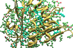
Photo: Serotonin Acetyltransferase, a protein involved in the conversion of serotonin to melatonin.


